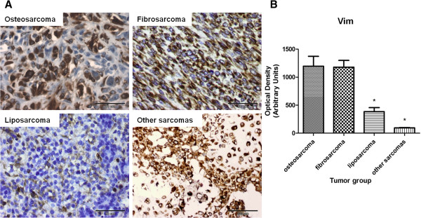
Figure 5.

Vimentin (Vim) expression in canine mammary sarcomas of various histological type. (A) Representative images of CMSs obtained under the Olympus BX41 microscope. Positive staining for Vim is observed as brown precipitate in the cytoplasm of neoplastic cells. The EnVision + System-HRP detection system was used, and the signal was visualized with chromogen 3,3-diaminobenzidine 3-3' (DAB). (B) The graph represents integrated optical density (IOD) of vimentin-positive cells in canine mammary sarcomas. The colorimetric intensity of IHC-stained antigen spots was determined in a computer-assisted image analyzer (Olympus Microimage™ Image Analysis version 4.0 for Windows, USA), and the color intensity of the antigen spot is expressed as mean pixel optical density on a 1–256 scale. The results are presented as the mean (±SEM) from all tumors in each group. Data was processed in Prism 5.00 software (GraphPad Software, California, USA) using one-way ANOVA and Tukey's HSD post-hoc test. P-values <0.05 (*) were regarded as significant and marked with an asterisk (*).
