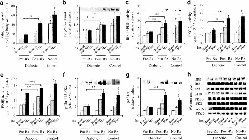
Fig. 1.
Effects of combined thiaziolidinedione–metformin treatment (Rx) on sub-maximal (Submax) and maximally (Max) effective insulin (Ins) on whole-body glucose disposal rates (a), phosphotyrosine content (reflective of activation) of the IR β-subunit (b), IRS-I/PI3K activity (c), atypical PKC-ζ/τ activity (d), PKBβ/Akt2 activity (e), phosphorylation of thrconinc-473 in total PKB (f) and phosphorylation of the PKB/Akt substrate AS160 (g) in muscles of type 2 diabetic participants. Basal, non-insulin-stimulated in clamp values. Also shown in each panel are basal and insulin-treated values of non-diabetic controls (Control) assayed in parallel with samples of diabetic participants. Values are mean±SE of five determinations for diabetic participants and four to five determinations for controls. ANOVA for effects of thiazolidinedione–metformin treatment in diabetic participants: *p < 0.05, **p < 0.01 and ***p < 0.001. Unless indicated otherwise, differences between treatment and pre-treatment values were not significant. †p < 0.05 for differences between mean values of control and untreated diabetic participants. Insert (c) shows representative autoradiogram of PI3K reaction product, PI3-PO4. Inserts (b, f, g) show representative immunoblots of phosphorylated proteins in western blot analyses, h Western blot analyses of: total insulin receptor β-subunit (tIRβ); IRS1; p85 subunit of PI3K (p85); p110 subunit of PI3K (p110); PKBβ; total PKBα/β (tPKB); total AS160 (tASl60); and total PKC zeta plus iota (tPKC-ζ/τ). Note that: (1) samples within immunoblots routinely contained (or were prepared from) equal amounts of lysate protein; and (2) except for aPKC, which is diminished in muscles of type 2 diabetic participants, no significant changes were seen in levels of indicated signalling proteins in muscles of diabetic vs control participants or in diabetic muscles after vs before treatment. Accordingly, it may be inferred that the levels of phosphorylated proteins in immunoblots reflected alterations to the phosphorylation status of a given amount of the signalling protein, rather than a change in the amount of signalling protein

