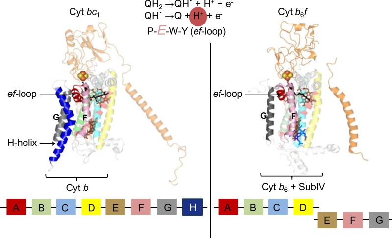
Figure 6. Organization of the Qp-site in cyochrome bc complexes.
The ef-loop bears the conserved PEWY sequence, whose Glu residue is involved in the second deprotonation reaction (highlighted in reaction sequence) of the substrate within the Qp-site. Left panel: In the cyt bc1 complex (PDB ID 3CX5), the ef-loop is inserted between the F and G transmembrane helices of the 8 helix cyt b polypeptide (shown in block diagram at the bottom). The space between the F and G TMH is stabilized by the H TMH of cyt b. Right panel: In the cyt b6f complex (PDB ID 2E74), the ‘H’ TMH is absent from the subIV polypeptide (shown as block diagram). This niche is occupied by a lipid and a chlorophyll-a molecule.

