
Figure 2.
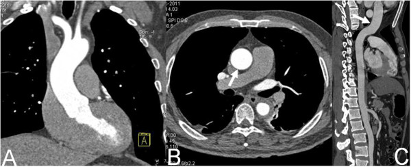
Dual-source CT imaging with high-pitch mode. Coronal MPR image (A, white arrow), axial original image (B, white arrow) and CPR image (C) show the wall of the entire ascending aorta is smooth without pulsation artifact.
Official websites use .gov
A
.gov website belongs to an official
government organization in the United States.
Secure .gov websites use HTTPS
A lock (
) or https:// means you've safely
connected to the .gov website. Share sensitive
information only on official, secure websites.
Dual-source CT imaging with high-pitch mode. Coronal MPR image (A, white arrow), axial original image (B, white arrow) and CPR image (C) show the wall of the entire ascending aorta is smooth without pulsation artifact.