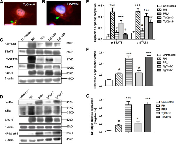
Figure 6.
TgCtwh3 and TgCtWh6 induced activation of macrophages through different signal pathway. (A and B) HeLa cells were infected with TgCtwh6 (A) or TgCtwh3 (B) for 19 hr, fixed, and stained with anti-NF-κB p65 (red), anti-Toxoplasma gondii p30-FITC (green), and Hoechst dye (blue). Arrow shows tachyzoites. This experiment has been done five times with similar results. (C and D) Total lysates were subjected to Western blotting using corresponding antibodies (for STAT6, pY-STAT6, STAT3, p-STAT3, IκBα, p-IκBα and SAG-1) or nuclear lysates (for NF-κB p65 subunit). The lysates were collected from BMMφs infected with either RH or PRU or TgCtwh3 or TgCtwh6 or left uninfected (2:1 ratio of tachyzoites to cells) at 24 h post-infection. (E and F) The fraction of phosphorylation was displayed. From the total STAT6, STAT3 or IκBα blot, the fraction of phosphorylated STAT6, STAT3 or IκBα was determined by comparing the intensity of the upper band (phosphorylated form) to the total intensity of the lower and upper band. Bars represent means ± SD from three independent experiments (#p>0.05, *p<0.05, ***p<0.001, either RH- or PRU- or TgCtwh3- or TgCtwh6-infected cells vs uninfected cells). (G) The nuclear relative expression of NF-κB p65 to nuclear actin using densitometry is also shown. Bars represent means ± SD (#p>0.05, *p<0.05, ***p<0.001). This experiment was repeated three times with similar results.

