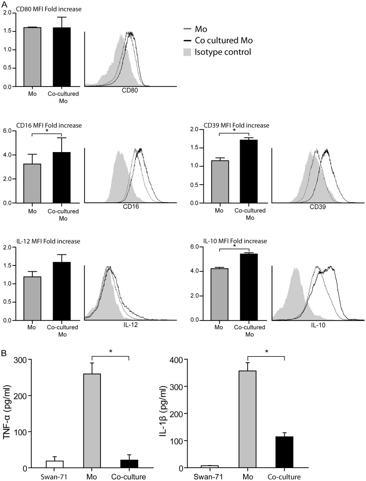
Figure 1. Trophoblast cells induce and alternative profile on maternal CD14+ cells.
Maternal monocytes purified by magnetic beads were cultured or not with Swan-71 cells at 70% of confluence in a 24 well flat-bottom plate. After 24 hours of culture, cells were recovered by TrypLE treatment and CD80, CD16, CD39, IL-12 and IL-10 were quantified by FACS on CD14+ gate (A). Results are expressed as fold increase of the MFI of the marker of interest relative to its isotype control (mean ± SEM) (*p<0.05, Wilcoxon test). Figure also shows a representative histogram of 3 independent experiments. At the same time, supernatants were recovered and TNF-α and IL-1β were quantified by ELISA (B). Results are expressed as mean ± SEM pg/ml from 3 independent experiments (*p<0.05, Wilcoxon test).

