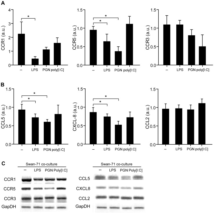
Figure 4. Trophoblast cells primed with PAMPs stimuli condition chemokine and chemokine receptor expression on maternal monocytes.
Swan 71 cell line at 70% of confluence were cultured in a polystyrene plate in complete DMEM 10% FCS in the absence or presence of LPS (10 µg/ml), PGN (10 µg/ml), or poly [I:C] (10 µg/ml) and 0.4 um pore-insert were added containing CD14+ cells. After 24 hours, cells were recovered from the upper compartments and the expression of (A) CCR1, CCR5 and CCR3; (B) CCL5, CXCL8, and CCL2 were evaluated by RT-PCR and normalized to GAPDH expression. Results are expressed as arbitrary units (a.u) (mean ± SEM) from 6 independent experiments using different fertile women (*p<0.05 Friedman test followed by Dunn’s post-test). (C) Representative amplification bands from 6 independent experiments.

