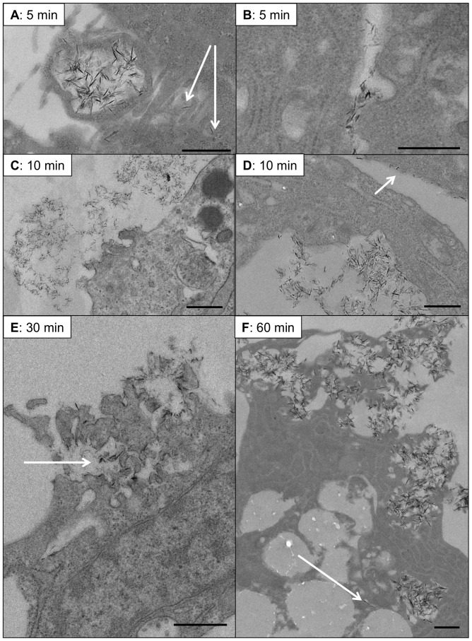
Figure 5. Ultrastructural analysis of CaP particle-exposure to VSMCs.
VSMCs were incubated with CaP particles (12.5 µg/mL) for specific times (as indicated) before fixing and processing for TEM analysis. A. Evidence for macropinocytosis of clusters of CaP particles was often observed after 5 minutes of particle exposure and uptake of individual particles was also seen at this early timepoint (indicated by arrows in A). Clathrin-like pits were also often observed after CaP particle exposure (B). After 10 minutes of CaP particle exposure, plasma membrane damage was observed in association with membrane protrusions (C) or ingression (D). Discrete CaP particles were also seen aligning at the plasma membrane surface (arrow in D). After 30 minutes, areas of plasma membrane damage were observed and these areas contained electron-dense particles (indicated by arrow in E). B. At 60 minutes, intracellular particle accumulation in clusters or isolated particles (indicated by arrow) were observed and large areas of plasma membrane rupture (F). Bar = 500 nm.

