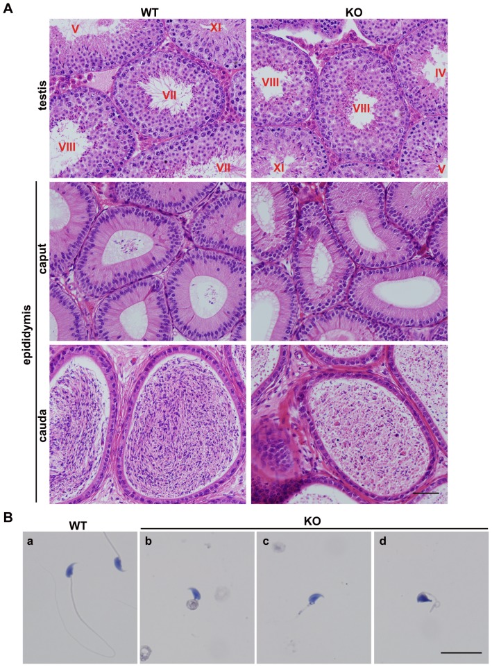
Figure 4. Iqcg was required for sperm flagellum formation in mice.
(A) H&E staining of WT and Iqcg KO testis and epididymis sections. In the WT testis, the lumens of seminiferous tubules of stage VII-VIII were filled with flagella. However, in the KO mice, no visible flagella could be found. The caput of WT epididymis displayed some spermatozoa in the lumens, but no spermatozoa were observed in the KO caput epididymis. WT cauda epididymis were filled with normal spermatozoa with long tails. However, the KO cauda epididymis only contained malformed spermatozoa and degenerated cell debris. The stages of seminiferous epithelial cycle were denoted by the Roman numerals. Scale bar = 50 µm. (B) H&E staining of WT and Iqcg KO spermatozoa on slides. WT spermatozoa showed a thin and long flagellum (a). Most KO spermatozoa showed an extremely short tail and were often connected by a mass of cytoplasm (b). Some KO spermatozoa only showed a short tail (c). A part of KO spermatozoa also displayed nuclear shaping defects (d). Scale bar = 20 µm.

