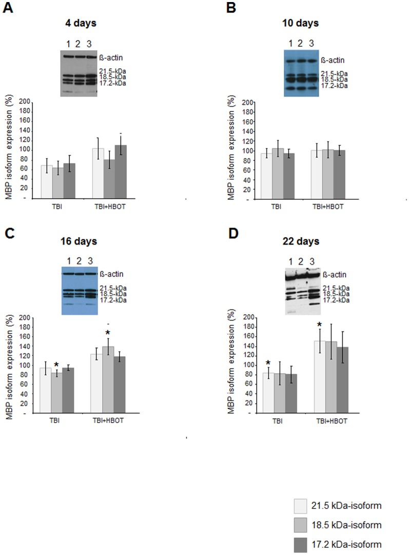
Figure 4. Western blot analysis of time-dependent changes in myelin basic protein isoform expression in the ipsilateral cortex following traumatic brain injury and HBO treatment.
A-D Myelin basic protein isoform expression at days 4, 11, 16 and 22 as percentage of expression in sham controls and representative western blot gels. Light grey bars: expression of isoform 21.5-kDa, grey bar: expression of isoform 18.5-kDa; dark grey bar: expression of isoform 17.2-kDa isoform; insert:, 1: sham, 2: brain injured animals, 3: injured and HBO-treated animals; western blot analysis was repeated at least twice per animal sample; n≥4 for each group. *p<0.05, HBO treated vs untreated injured animals; # p<0.05 as compared to sham controls. Error bars represent ± standard error means.

