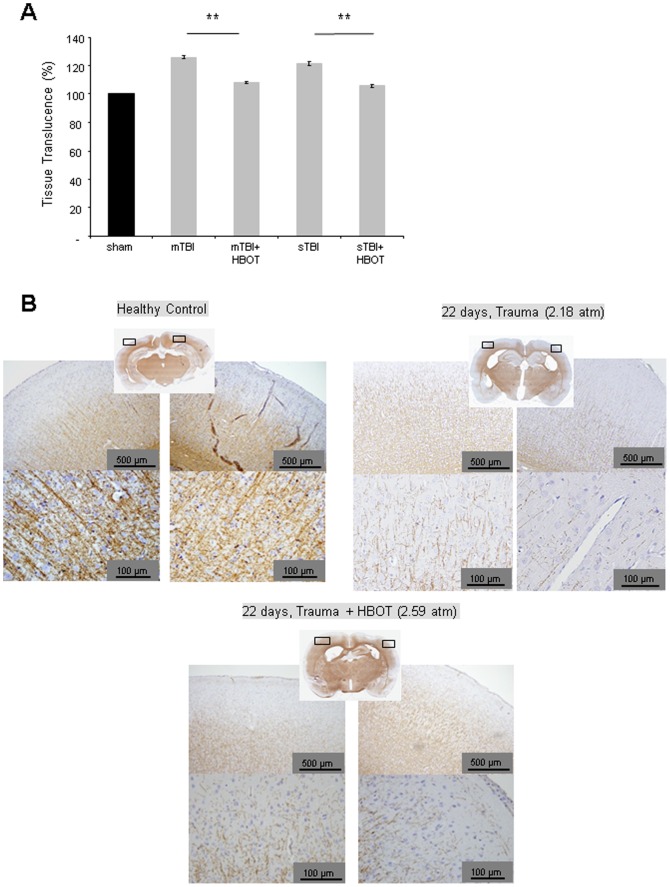
Figure 5. Modulation of myelin in the ipsilateral cortex at day 22 following induction of traumatic brain injury and HBO treatment.
A. Quantitative analysis of myelin stained by Luxol Fast Blue. The ipsilateral hemisphere was analysed as a whole in order to avoid bias. Representation of the % tissue translucence of the ipsilateral hemisphere as compared to sham controls; a minimum of 4 successive brain slices per animal were analysed, *p<0.05, HBO-treated versus untreated animals; B. Exemplification of proteolipid protein (PLP) staining at day 22 following induction of traumatic brain injury and HBO treatment; number of animals see Table 1. Error bars represent ± standard error means.

