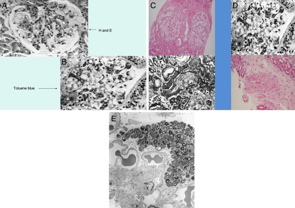
Figure 2.
Histological diagnosis of Fabry disease. (A) Haematoxylin and eosin stain of tissue from a patient with Fabry disease showing lesions that could be confused with other conditions in which foam cells are present; (B) Semi-thin section stained with toluene blue (the preferred method for diagnosing Fabry disease on renal histology); (C) deposits in the tubules that can give rise to mild renal tubular disorders; (D) a small artery with large degrees of narrowing due to Gb3 deposits; (E) ultrastructural image (electron microscopy) of the glomerulus in Fabry disease shows lamellated lipid inclusions (zebra bodies) within the podocyte cytoplasm (×6,000; stain: UrPb). Reproduced with the permission of Dr. Kostas Giannakakis (Laboratory of Ultrastructural Pathology, Rome La Sapienza University, Rome, Italy). E, eosin; H, haematoxylin; UrPb, uranyl lead.

