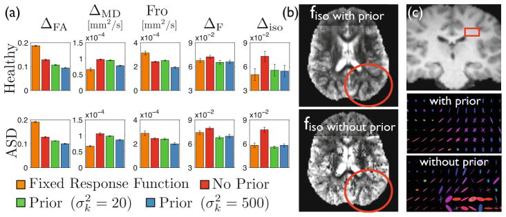
Fig. 3.

(a) Incorporating prior knowledge significantly improves the quality of the model estimation for all five metrics and for both healthy controls and ASD patients. This improvement implies that (b) the extracellular water fraction can be visualized with more contrast and less noise in smaller details of the white matter up to its boundary with the grey matter, and (c) properties of the fascicles in crossing areas (shown is the corona radiata) are better represented and do not suffer the arbitrary choice of a model from Equation (3).
