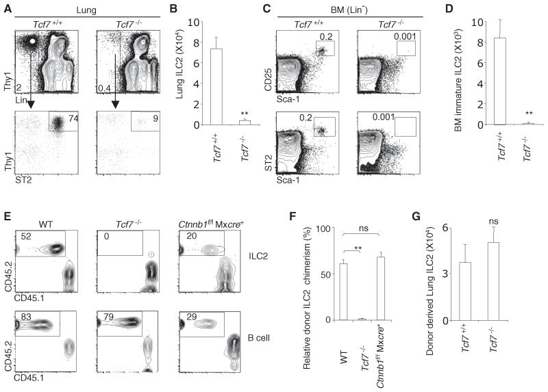
Figure 1. TCF-1 Is Required for ILC2 Development.
(A) Flow plots show a representative analysis of ILC2 in the lungs of Tcf7+/+ and Tcf7−/− mice. Plots were gated on CD45+ lung cells.
(B) Bar graph shows cellularity of lung ILC2 in Tcf7+/+ and Tcf7−/− mice. Data are from eight mice per group (error bars are SEM).
(C) Flow plots show a representative analysis of bone marrow immature ILC2 in Tcf7+/+ and Tcf7−/− mice. Plots were gated on Lin− cells.
(D) Bar graph shows cellularity of bone marrow immature ILC2. Data are from three or four mice per group (error bars are SEM).
(E) We mixed 5,000 LSK cells from WT, Tcf7−/− and poly(I:C)-treated Ctnnb1(encoding β-catenin)f/f Mxcre mice (CD45.2) with 2,500 WT competitor LSK cells (CD45.1) and together injected them into lethally irradiated (950 rads) WT recipients (CD45.1). Reconstitution of lung ILC2 and splenic B cells was examined at 4 weeks posttransplant. Deletion of Ctnnb1 in LSK cells was confirmed by PCR (data not shown).
(F) Bar graph shows relative ILC chimerism normalized to splenic B cells. Results are from two independent experiments; five mice per group (error bars are SEM).
(G) We intravenously transferred 5,000 LSK cells from WT mice (CD45.1) into lethally irradiated Tcf7+/+ and Tcf7−/− mice (CD45.2). Development of lung ILC2 was examined at 4 weeks posttransplant. Data are from two independent experiments; three mice per group (error bars are SEM; ns, not significant). **p < 0.05. See also Figure S1.

