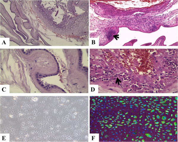
Figure 1.
Histologic images: A – normal placental membranes, HE (Hematoxylin & Eosin, the routine staining) x 100; B – chorioamnionitis with perivascular inflammatory infiltrate (arrow head), HE x 100; C – normal amnion, HE x 400; D – chorioamnionitis with perivascular inflammatory infiltrate (arrow head), HE x 400; E – HEAC in culture, phase contrast x 100; F – CX3CR1 immunostained in HAEC culture (image digitally transformed for morphometric purposes), x 200.

