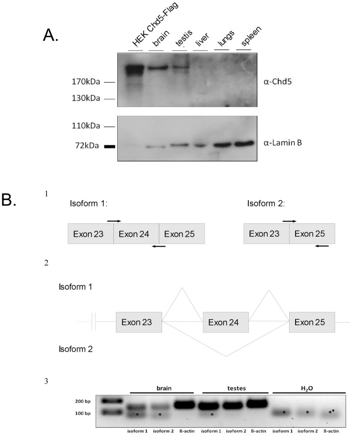
Figure 2. CHD5 protein is expressed in brain and testis.
(A) CHD5 protein is expressed in mouse brain and testis. Western blot was performed with CHD5 antibody on lysates from different mouse tissues. Extracts from HEK cells transiently transfected with CHD5-Flag served as a positive control. Lamin B was used as a loading control. (B) Two different CHD5 isoforms are expressed in mouse brain and testis. Panel 1: schematic representation of the location of the PCR primers used to detect CHD5 cDNA of isoforms 1 and 2. Panel 2: schematic representation of part of the CHD5 gene structure showing the two different putative splicing variants. Exons 23, 24 and 25 are indicated. Horizontal lines are introns. Diagonal lines indicate splicing. Panel 3: RT-PCR detecting expression of different CHD5 isoforms in mouse testis and brain. H2O: negative control. Asterisk: primer dimers.

