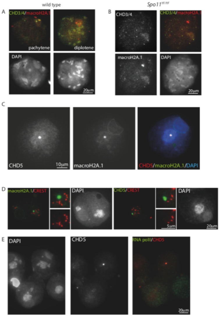
Figure 8. macroH2A colocalizes with CHD3/4 in spermatocytes and with CHD5 in spermatids.
(A,B) Spread wild type (A) and Spo11 null (Spo11YF/YF) (B) spermatocyte nuclei stained for MacroH2A.1 and CHD3/4. Colocalisation on the PAR and the X centromere is observed in pachytene, but not in diplotene nuclei of wild type spermatocytes. In the absence of functional SPO11, some zygotene-like nuclei show enrichment of both CHD4 and macroH2A.1 in the pseudo XY body. (C) Spread spermatid nucleus stained for CHD5, and MacroH2A, colocalisation of the CHD5/MacroH2A focus in the chromocenter is clear in the merge with DAPI (right image). (D) Spread spermatid nuclei stained with macroH2A (green) and CREST (red, left panel) or with CHD5 and CREST (right panel). The enlargements in the middle of each panel clearly show that the macroH2A.1/CHD5 spot in the chromocenter does not colocalise with CREST marking of the centromeres. (E) Spread spermatid nuclei costained for CHD5 and RNA polymerase II. Both high and low levels of RNA polymerase II can be observed in nuclei that express CHD5.

