
Figure 1. Activation of Akt pathway by pemetrexed.
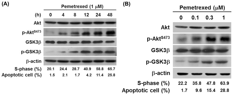
(A) Akt activation in a time course dependent manner. Human lung adenocarcinoma A549 cells were treated without or with 1 µM pemetrexed for 0, 4, 8, 12, 24, and 48 h, and then cell lysates were isolated. (B) Akt activation occurred in a concentration dependent manner. A549 cells were treated with 0, 0.1, 0.3 and 1 µM pemetrexed for 48 h. After treatment, the levels of total Akt, phosphorylated Akt, total GSK3β, and phosphorylated GSK3β were examined by Western blot analysis. β-Actin was used as an internal loading control. The proportion of S-phase population and apoptotic cells was determined as described in the Materials and Methods section.