
Figure 4. Akt translocates to the nucleus during pemetrexed-induced cell death.
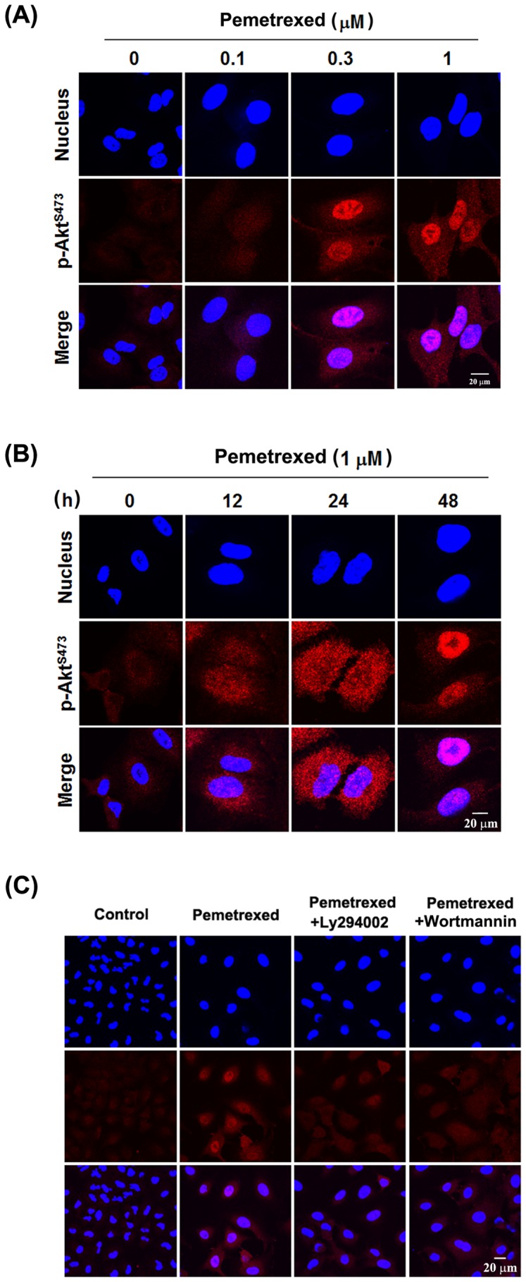
(A) A549 cells were treated with 0, 0.1, 0.3, and 1 µM pemetrexed for 48 h, and (B) A549 cells were treated with 1 µM pemetrexed for 0, 12, 24, and 48 h. After treatment, the subcellular distribution of p-AktS473 was detected by confocal microscopy after immunostaining with anti-phospho-AktS473 and Rhodamine-conjugated secondary antibody. Hoechst 33342 was used to counter stain nuclei, and the images were overlaid to determine the Akt localization within the cell. (C) Inhibiton of Akt activation by Ly294002 and wortmannin blocked Akt nuclear accumulation. A549 cells were pretreated with 10 µM Ly294002 or 3 µM wortmannin for 2 h, and then 1 µM pemetrexed was added for another 48 h. The subcellular distribution of p-AktS473 was detected by immunocytochemistry.