
Figure 6. Activation of Akt and Cdk2 are occurred in pemetrexed-treated H1299 cells.
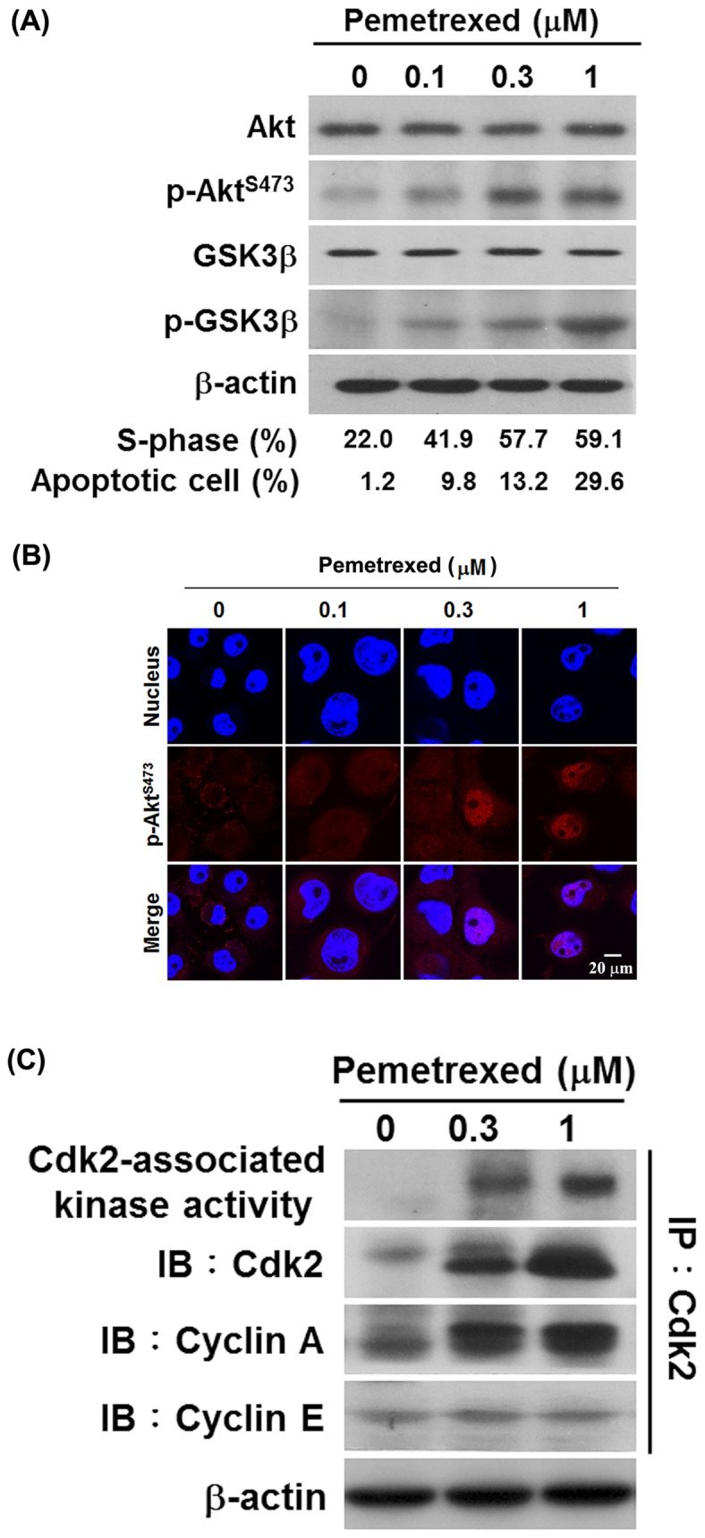
(A) Pemetrexed stimulates Akt pathway activation. H1299 cells were treated with 0, 0.1, 0.3 and 1 µM pemetrexed for 48 h. After treatment, the levels of total Akt, phosphorylated Akt, total GSK3β, and phosphorylated GSK3β were examined by Western blot analysis. β-Actin was used as an internal loading control. The proportion of S-phase population and apoptotic cells were determined as described in the Materials and Methods section. (B) Nuclear accumulation of Akt occurred in pemetrexed-treated H1299 cells. Cells were treated with 0, 0.1, 0.3, and 1 µM pemetrexed for 48 h, the subcellular distribution of p-AktS473 was detected by confocal microscopy after immunostaining with anti-phospho-AktS473. Hoechst 33342 was used to counterstain nuclei. (C) Pemetrexed activated Cdk2/Cyclin A-associated kinase in H1299cells. H1299 cells were treated with 0, 0.3, and 1 µM pemetrexed for 24 h, and then protein lysates were isolated. The Cdk2 kinase activity and the levels of Cdk2, Cyclin A and Cyclin E were determined.