
Figure 1.
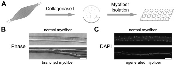
Isolation of single myofibers. (A) Muscles were digested with collagenase type I, and single myofibers were isolated. (B) A branched myofiber contains one or more small myotubes contiguous with the parent myofiber. Bar = 100 μm. (C) Myofibers were stained with 4′,6-diamidino-2-phenylindole (DAPI) to visualize nuclei. A myofiber with at least four centrally nuclei located in a row was considered regenerated. Bar = 100 μm.