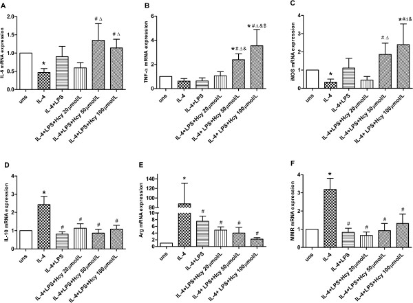
Figure 3.
Effects of Hcy and LPS on mRNA expression in M2 macrophage. (A) IL-6, (B) TNF-α, (C) iNOS, (D) IL-10, (E) Arg-1 and (F) MMR mRNA expression were assessed by real-time PCR. Gene expression is represented as fold-change compared to unstimulated macrophages. Data are representative of three independent experiments. (uns: unstimulated macrophages; Arg stands for Arg-1; *p < 0.05 vs uns; #p < 0.05 vs IL-4; Δp < 0.05 vs LPS; &p < 0.05 vs LPS + Hcy 20 μmol/L; $p < 0.05 vs LPS + Hcy 50 μmol/L).

