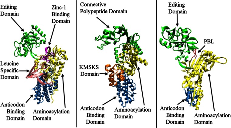
Fig. 1.
Cartoon representation of the 3D structure of the monomeric form of a Tt LeuRS (pdb code: 1H3N, residues 1–814); b Ec MetRS (pdb code: 1qqt, residues 3–548); c Ef ProRS (pdb code: 2J3M, residues 19–567). The structural domains are colored as follows: a green, the editing domain (ED; residues 224–417); purple, zinc-1 binding domain (ZBD; residues 154–189); yellow, aminoacylation domain (residues 1–153, 190–223 and 638–642); pink, leucine-specific domain (LSD; residues 577–634); and blue, the anticodon binding domain (residues 635–814); b yellow, aminoacylation domain (residues 1–96, 252–323); green, connective polypeptide (CP) domain (residues 97–251); orange, KMSKS domain (324–384 and 536–547), and blue, the anticodon binding domain (residues 385–535); c green, editing domain (residues 224–407), red, the proline-binding loop (residues, 199–206); red, aminoacylation domain (residues 1–223, 408–505), and blue, the anticodon binding domain (residues 506–567)

