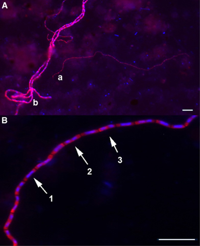
Figure 3.
(A) Cable bacteria in incubated marine sediment targeted by the DSB706 probe, showing different phenotypes of cable bacteria with (a) 0.7 μm (t21) and (b) 1.2 μm (t21) diameters. (B) Different stages of cell division were detected in cells along the multicellular cables (1) daughter chromosomes located side by side, (2) cell division is initiated in the middle, (3) daughter chromosomes are located in the middle of the daughter cells. Scale bars, 10 μm.

