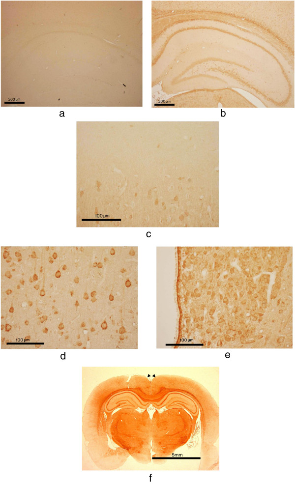
Figure 4.
D-AIB AT immunohistochemistry in 8-week male rat brains. Figure 4a High magnification of hippocampal region with pre-immune rabbit serum, Figure 4b; with rabbit anti D-AIB AT antibody are shown. Higher magnification of outer layer of cerebral cortex (Figure 4c), inner layer of the cerebral cortex (Figure 4d), and brain stem (Figure 4e) are shown with anti D-AIB antibody. Figure 4f. Whole rat brains with coronal sections (arrowheads indicates retrosplenial cortex) are shown with anti D-AIB antibody.

