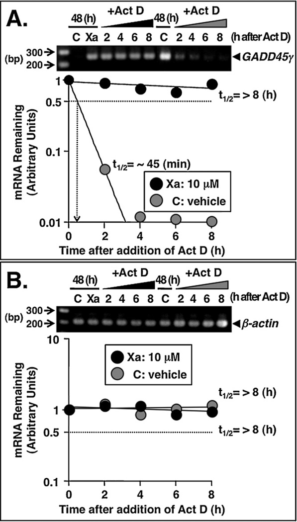
Fig. 7.
(−)-Xanthatin stabilizes GADD45γ mRNA. (A and B) Effects of (−)-xanthatin or vehicle on the mRNA stability of GADD45γ and β-actin in MDA-MB-231 cells. MDA-MB-231 cells were treated for 48 h with 10 µM (−)-xanthatin (Xa) or vehicle (C), and subsequently, the cells were exposed to the transcriptional inhibitor, actinomycin D (Act D, 4 µg/mL) for 2, 4, 6, or 8 h. The Act D concentration used was determined based on both efficacy and lack of toxicity following dose-response experiments. After the respective Act D exposures, total cellular RNA was isolated and RT-PCR analyses were performed as described in Section 2. β-Actin was also used as an RNA internal control. (A) Semi-logarithmic plot of the decay of GADD45γ mRNA is shown. The number of optimized PCR cycles used for the amplification of GADD45γ was 33. Due to the very low expression levels of GADD45γ, for the determination of half-life of GADD45γ mRNA after ‘vehicle exposure (indicated as C)’, the number of optimized PCR cycles used for the amplification of GADD45γ was 42. A 100 bp DNA ladder marker was also loaded. (B) Semi-logarithmic plot of the decay of β-actin mRNA is shown. The number of optimized PCR cycles used for the amplification of β-actin was 29. The mRNA decay plots and calculation of the mRNA half-life were performed according to reported methods (Rishi et al., 1999). A 100 bp DNA ladder marker was also loaded.

