Figure 5.
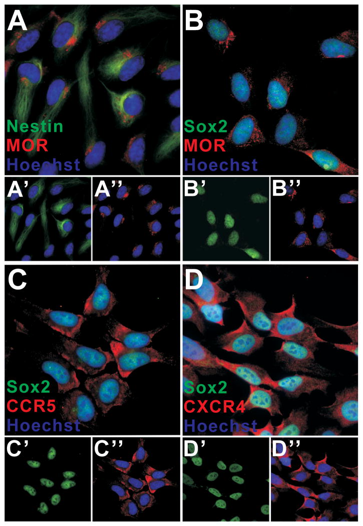
Immunohistochemical characterization of hNPCs. Double-labeling confirmed that 95% of hNPC were nestin+ and Sox2+; 95% also showed immunoreactivity for MOR (A, B). Individual precursor markers are separated in the lower panels (A′: nestin (green) and nuclei (blue); A″: MOR (red) and nuclei (blue); B′: Sox2 (green); B″: MOR (red) and nuclei (blue)). A similar percentage of hNPC express CCR5 (C) and CXCR4 (D). As above, lower panels show individual markers (C′: Sox2 (green); C″: CCR5 (red) and nuclei (blue); D′: Sox2 (green); D″: CXCR4 (red) and nuclei (blue)). The presence of CCR5 and CXCR4 on hNPCs suggests that effectors such as gp120, β-chemokines (RANTES/CCL5, MIP-1α/CCL3, MIP-1β/CCL4), and the α-chemokine (SDF-1/CXCL12), released in response to Tat or contained in viral supernatant, might affect hNPC behavior.

