Figure 7.
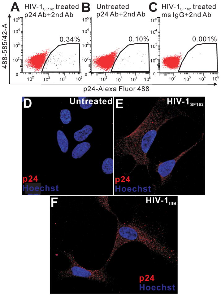
Detection of p24 on hNPCs by FACS and confocal microscopy. p24 immunolabeling was assessed on 1 × 10⁴, 7-AAD+ cells using a single laser (488 nm) and two color fluorescence emission (585/42 nm band pass; 530/30 hm band pass) FACS approach (A–C). A very small percentage (0.33%) of hNPCs exposed to supernatant from HIVSF162-infected monocytes were p24+ (A). Labeling indices in control groups, including untreated hNPCs immunolabeled for p24 (B) and HIV-treated hNPCs exposed to control IgG and second antibody (C), were 0.1% and 0.001%, respectively, suggesting that some p24 labeling was specifically related to antigen expression. Autofluorescence (585 > 530 signal) was not detected in any sample. FACS diagrams are representative of n=3 experiments. p24 signal was not detected in individual untreated hNPCs (D). p24 immunoreactivity was found within cytoplasm of cells exposed to both HIV-1SF162 (E) and HIV-1IIIB supernatant (F). Images show 0.32 μM optical sections through the nuclear region. Note the absence of p24 immunolabeling in nuclei.

