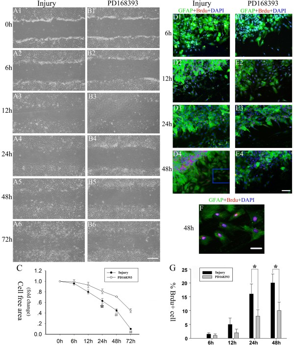
Figure 2.
PD168393 treatment slowed down scratch injury-induced reactive astrogliosis. Astrocytes were scratched and incubated in the absence or with treatment of 40 μM PD168393. (A1-B1), (A2-B2), (A3-B3), (A4-B4), (A5-B5) and (A6-B6) are paired images taken from control 0, 6, 12, 24, 48 and 72 hour groups after scratch injury, respectively. Scale bars = 100 μm in B6 (applies to A1-B6). Graphical representation of remaining cell-free area quantification over 72 hours after scratch injury (C). Data are expressed as fold change compared to control group (0 hours). Values are expressed means ± SD (n = 5). Significant difference between time points was observed (*P < 0.05, #P < 0.01). Immunostaining for Brdu, glial fibrillary acid protein (GFAP) and 4,6-diamidino-2-phenylindole (DAPI) at 6, 12, 24 and 48 hours after scratch injury in the injury group (D1-D4) and PD168393 (40 μM) treatment group (E1-E4), respectively. Astrocytes were identified by GFAP immunostaining (green) and nuclei were identified by DAPI labeling (blue). Astrocytes in S phase were identified by BrdU labeling (red). Insets in panels (D4) of co-localization of phosphorylated epidermal growth factor receptor (pEGFR), GFAP and DAPI are shown at high magnification in (F). Scale bars = 100 μm in E4 (applies to D1-E4); scale bars = 20 μm in (F). Percentage of astroglial cells with BrdU labeling at different times after scratch injury (G). *P < 0.05.

