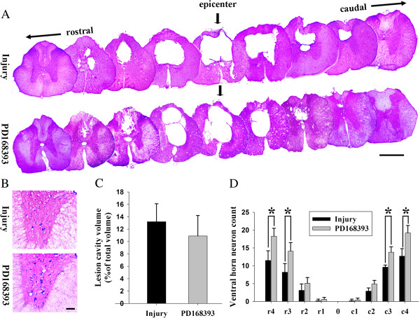
Figure 7.
PD168393 promoted survival of ventral horn (VH) motor neurons after spinal cord injury (SCI). Representative transverse cresyl violet eosin stained sections of four weeks post-SCI at the epicenter and in 1 mm increments rostral and caudal to the epicenter. Epicenter sections are indicated by arrows. Scale bar = 1 mm (A). Representative photomicrographs from four weeks post-SCI showing cresyl violet eosin stained VH neurons at 4 mm rostral to the injury epicenter. Scale bar = 50 μm (B). Graphical representation showing no statistically significant difference in lesion cavity volume following treatment (n = 5) (C). Comparison of VH neurons among different groups at various distances from the injury epicenter (0) as well as 1 to 4 mm rostral (r) and caudal (c) to it (D) (n = 5, *P < 0.05).

