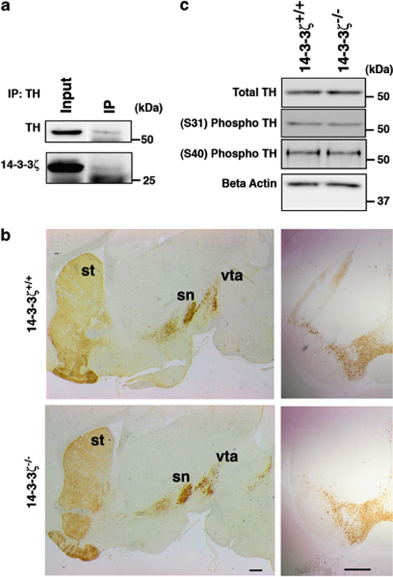
Figure 4.
Tyrosine hydroxylase (TH) is activated normally in 14-3-3ζ knockout (KO) mice. (a) TH was precipitated from P35 whole-brain lysates with anti-TH antibody. TH immunoprecipitates were probed with anti-TH and monoclonal antibodies against 14-3-3ζ (M6). (b) Sagittal sections and (i, ii) coronal sections (iii iv) show that the abundance of TH-positive dopaminergic neurons in the substantia nigra (sn) and ventral tegmental area (vta), and their projections to the striatum (st) are similar in 14-3-3ζ KO and wild-type (WT) mice. Scale bars=500 μm. (c) Western blot analysis of brain lysates shows that total TH, phospho serine 31 (Ser-31) and phospho Ser-40 are present at similar levels in 14-3-3ζ KO and WT mice. Load control used for quantitating western blots was α tubulin. A representative blot of all four samples is shown in this figure that is quantitated in Supplementary Figure S3.

