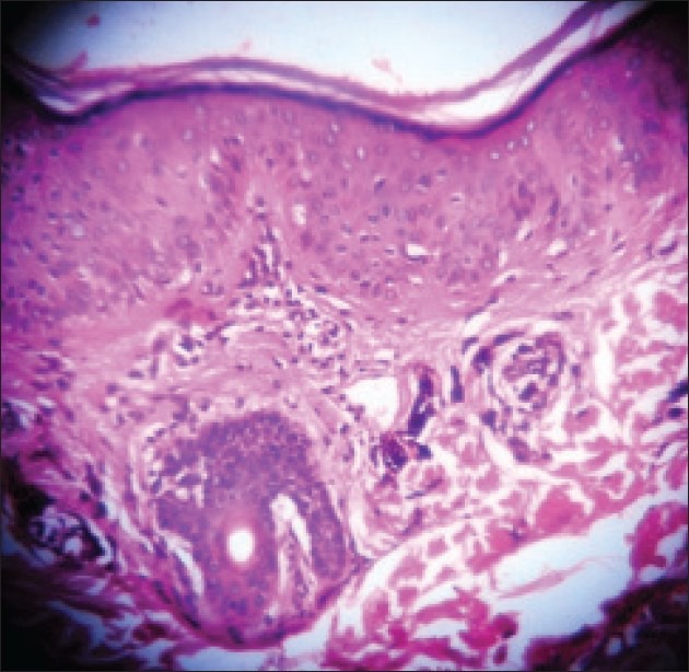
Figure 2.

Histopathological examination of the section showed epidermis with hyperkeratosis, extensive vacuolar change, and basal cell degeneration. There was melanin incontinence with numerous melanophages and perivascular and periadnexal chronic inflammation (H and E, ×10)
