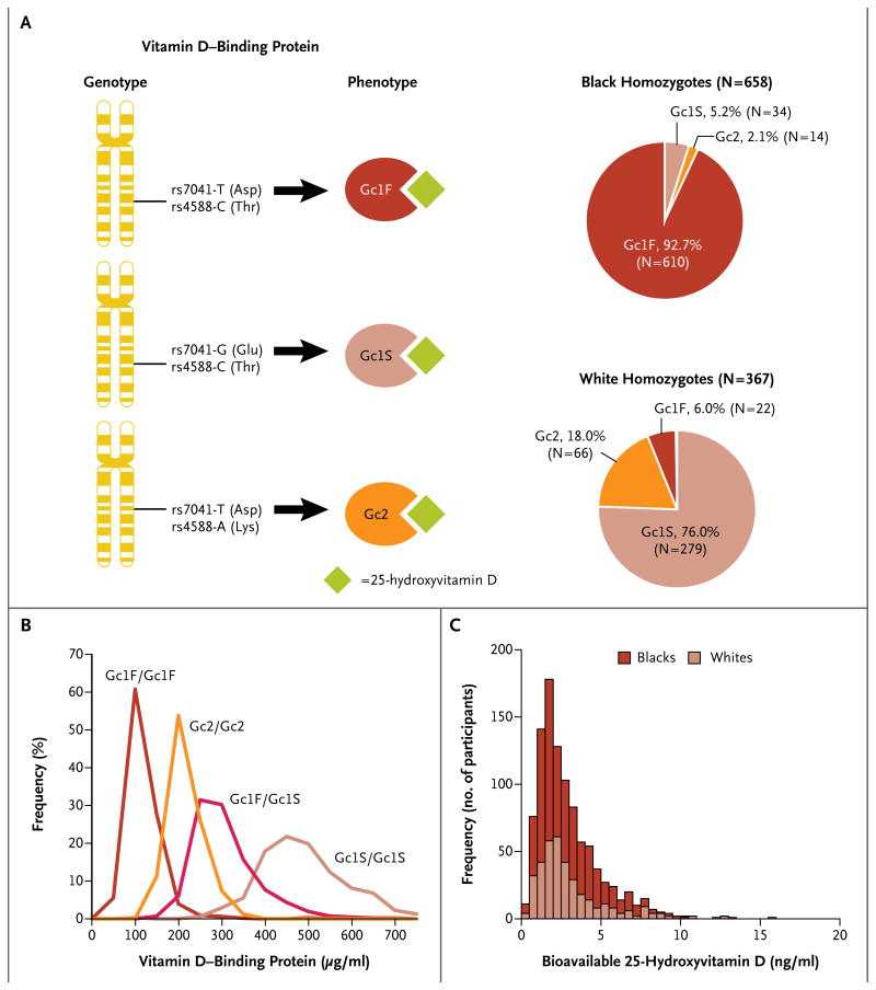
Figure 2. Variant Vitamin D–Binding Proteins and Bioavailable 25-Hydroxyvitamin D.
As shown in Panel A, unique combinations of the rs7041 and rs4588 polymorphisms produce amino acid changes resulting in variant vitamin D–binding proteins (left side of panel; Asp denotes aspartic acid, Glu glutamic acid, Lys lysine, and Thr threonine). The Gc1F phenotype was most common in black homozygotes, whereas the Gc1S phenotype was most common in white homozygotes (right side of panel). As shown in Panel B, levels of vitamin D–binding protein were lowest in Gc1F/Gc1F homozygotes (632 participants, 93±2 μg per milliliter), highest in Gc1S/Gc1S homozygotes (313 participants, 468±6 μg per milliliter), and intermediate in Gc2/Gc2 homozygotes (80 participants, 190±4 μg per milliliter). Plasma vitamin D–binding protein concentrations in Gc1F/Gc1S heterozygotes (413 participants, 285±4 μg per milliliter) were intermediate between those of Gc1F/Gc1F homozygotes and Gc1S/Gc1S homozygotes. These differences were significant (P<0.001 for all comparisons). Panel C shows a histogram representing stacked distributions. Among homozygous participants, levels of bioavailable 25-hydroxyvitamin D were similar in blacks and whites (2.9±0.1 ng per milliliter in blacks and 3.1±0.1 ng per milliliter in whites, P = 0.71).

