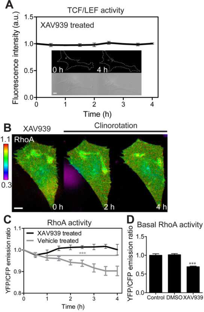
Fig. 4.
The role of β-catenin signaling on the RhoA activity by clinorotation. (A) TCF/LEF activity of XAV939-treated MC3T3-E1 cells under clinorotation. n = 6 cells. (B, C) RhoA activity of XAV939-treated MC3T3-E1 cells under clinorotation. RhoA activity failed to respond to simulated unloading. For statistical analysis of RhoA activity in vehicle (DMSO)-treated cells, the emission ratios were compared with those at time 0 (*** P < 0.001). n = 6 cells in both cases. (D) Basal RhoA activities of control cells, vehicle-treated cells, and XAV939-treated cells. n = 10 (Control); 6 (DMSO); 7 cells (XAV939). *** P < 0.001. Scale bars 10µm.

