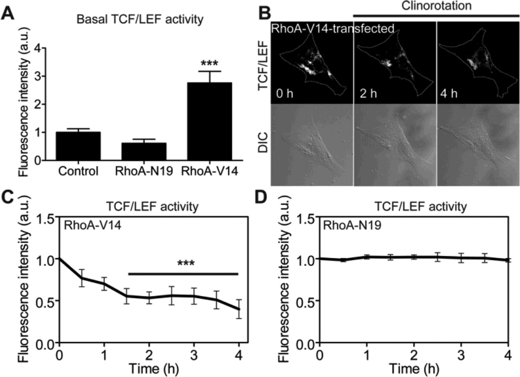
Fig. 5.
β-catenin signaling is rapidly, significantly reduced in MC3T3-E1 cells expressing constitutively active RhoA under clinorotation. (A) Basal TCF/LEF activities of control cells, and cells transfected with RhoA-N19 or RhoA-V14. RhoA-V14 data was compared with Control and RhoA-N19 data (*** P < 0.001). At least seven cells were analyzed. (B) β-catenin signaling was determined by monitoring the fluorescence intensity of the cells co-transfected with a TCF/LEF-GFP reporter and RhoA-V14. (C) Changes in TCF/LEF activity in response to clinorotation in MC3T3-E1 cells transfected with a constitutively active RhoA (RhoA-V14). n = 7 cells. For the statistical analysis, time course of TCF/LEF activity was compared with 0 hour (*** P < 0.001). (D) Changes in TCF/LEF activity in response to clinorotation in MC3T3-E1 cells transfected with a dominant negative RhoA (RhoA-N19). n = 7 cells.

