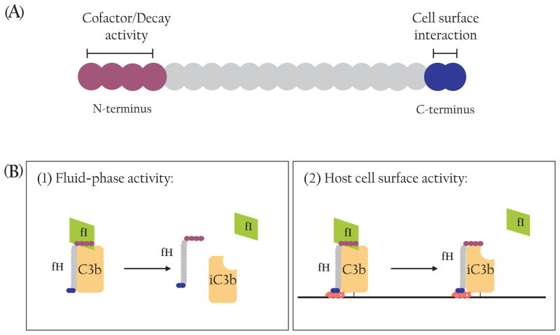
Fig. 3.
Structure and function of Factor H. (A) Schematic representation of fH functional domains. Each SCR is shown as a filled circle. The four N-terminal SCR contain cofactor and decay-accelerating activities while the two C-terminal SCR are primarily responsible for binding to the host cell surface. (B) Factor H is a unique complement regulator as it inhibits AP complement activity in the fluid-phase (1) as well as on the host cell surface (2). Only the cofactor activity of fH is illustrated here.

