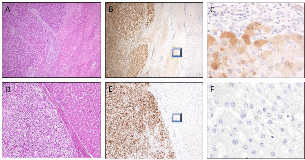
Figure 2.
AKR1B10 expression in hepatocellular carcinomas. Hepatocellular carcinoma (HCC) arising in a cirrhotic liver (A; H&E stain, 100X). Strong and diffuse AKR1B10 protein expression in HCC with faint cytoplasmic staining observed in the proliferating cells at the periphery of regenerative nodules (B, 100X and C, 600X). HCC in a non-cirrhotic liver (D; H&E stain, 200X) highlights strong and diffuse cytoplasmic and nuclear staining of AKR1B10 (E, 200X) and negligible AKR1B10 expression in the adjacent hepatic parenchyma (F, 600X).

