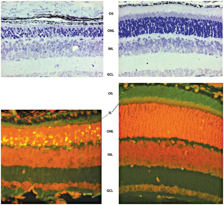
Figure 3.

Effect of TUDCA on rd10 mouse retinal morphology. Representative micrographs stained with toluidine blue showing significant preservation of photoreceptor nuclei in the outer nuclear layer (ONL) in TUDCA-treated eyes (top panel, right) compared with vehicle-treated eyes (top panel, left). Counts of photoreceptor nuclei were significantly greater in eyes with TUDCA treatment compared to vehicle only. Combined inner segment (IS) and outer segment (OS) length was similarly preserved, with no change in thickness of inner nuclear layer (INL) or ganglion cell layer (GCL). Fluorescence microscopy using a B-2A long pass emission fluorescence filter allows further observation of the preservation of photoreceptor IS and OS present in TUDCA-treated (bottom panel, right) vs vehicle-treated (bottom panel, left) retinal sections. TUNEL-positive nuclei (green/yellow signal) are seen to be abundant in vehicle-treated sections, but are rare in TUDCA-treated sections. TUDCA treatment provided significant preservation of photoreceptor nuclei number in the ONL. Treatment had no discernable effect on the INL or GCL.
Reprinted with permission from Boatright et al, 2006.32
