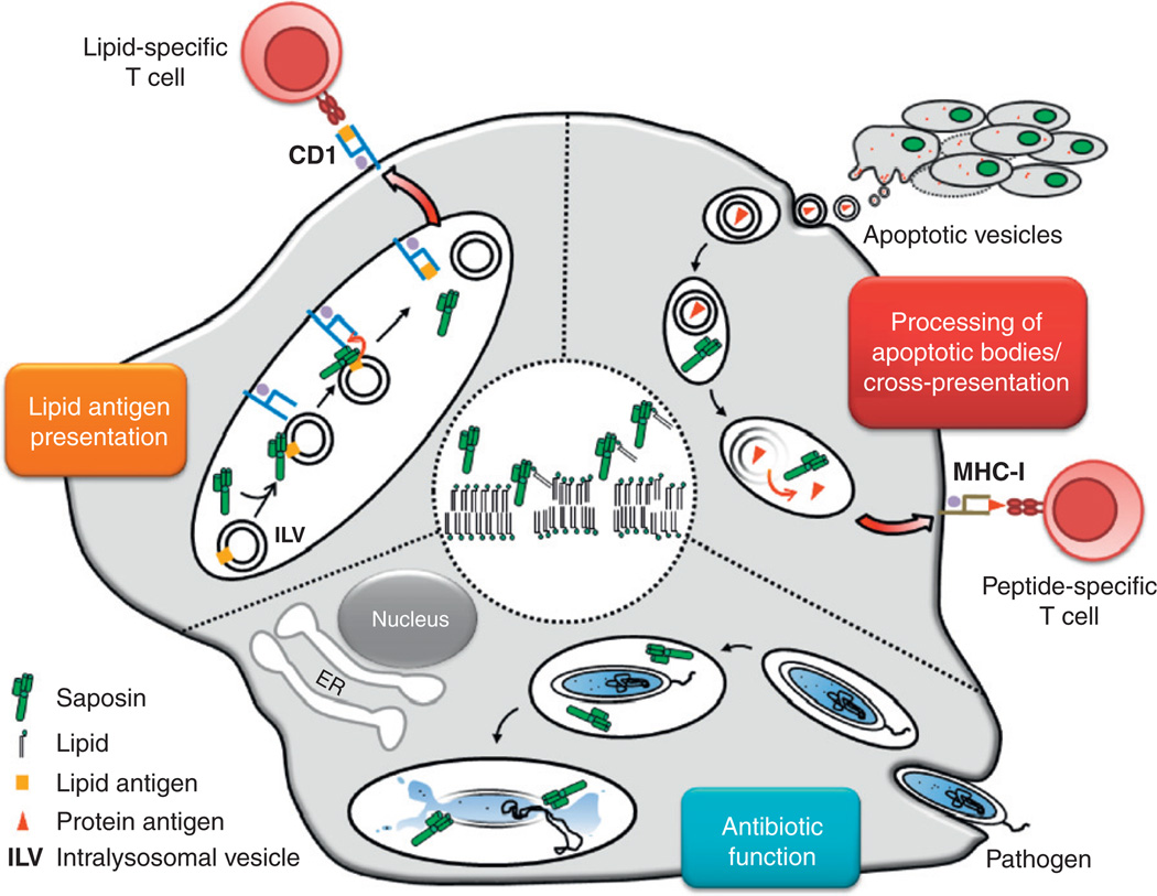
Figure 2.2.
Immunological functions of saposins. The central circle demonstrates the universal mechanism of saposin action on lipid bilayers. Accordingly, saposin molecules insert into lysosomal membranes to induce bilayer disintegration and lipid extraction. Further, the graph represents the three immunological consequences of this mechanism. (1) Lipid antigen presentation: In lysosomes of APCs, saposins interact with intralysosomal vesicles (ILVs) that contain lipid antigens. Subsequently, SAPs facilitate the loading of CD1 molecules with lipid antigens to activate CD1-restricted lipid-reactive T cells. (2) Processing of apoptotic bodies / cross-presentation: Apoptotic bodies derived from tumor or infected cells are engulfed by phagocytes such as DCs or macrophages. Protein antigens contained in apoptotic vesicles are liberated in lysosomes upon membrane disintegration induced by saposins. This leads to antigen delivery from apoptotic bodies for subsequent cross-presentation of antigens to MHC-I-restricted CD8+ T cells. (3) Antibiotic function: Pathogens like intracellular bacteria are phagocytosed by macrophages or DCs. At the acidic pH of lysosomes, saposins unfold their antimicrobial effects through direct attack on the membranes of pathogens.

