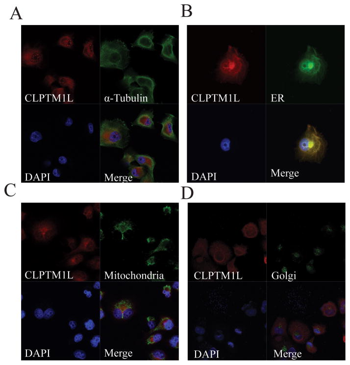
Figure 1. Localization of endogenous CLPTM1L in pancreatic cells and co-localization of CLPTM1L with cytoplasmic organelles.
(A) Localization of endogenous CLPTM1L in PANC-1 pancreatic cancer cells. Cytoplasmic and punctate nuclear, or nuclear membrane, staining is seen (red). Counter staining was performed for α-tubulin (green). (B) Co-localization of WT CLPTM1L (red) with the Endoplasmic Reticulum (ER) marker Calnexin (green) in PANC-1 cells stably transfected with WT CLPTM1L. Co-localization of WT CLPTM1L (red) with a mitochondrial marker (green) (C), or a Golgi marker (GM130 in green) (D), was not seen in PANC-1 cells stably transfected with WT CLPTM1L. Cells in all panels were counterstained with DAPI (blue).

