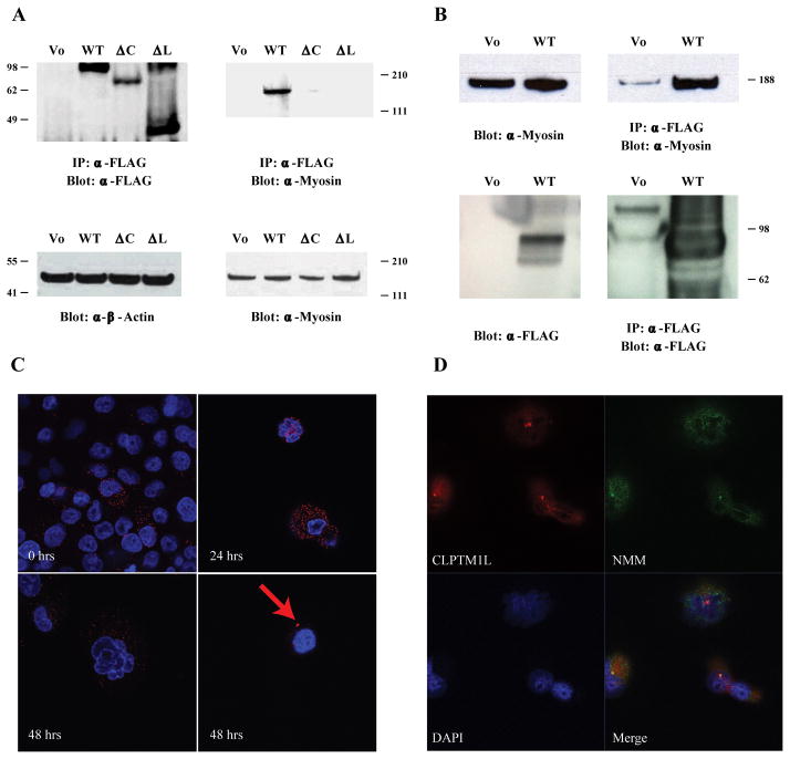
Figure 4. CLPTM1L and non-muscle Myosin II interact.
(A) Co-immunoprecipitation and western blot analysis showing robust interaction between WT N-FLAG CLPTM1L and non-muscle myosin II (Myosin) in stably transfected PANC-1 cells (upper right panel). Little or no interaction was seen with the two deletion mutants of CLPTM1L (upper left panel). β-Actin protein levels in cell lysates are shown as a loading control (lower left panel) and non-muscle myosin II (Myosin) protein levels are shown by western blot (lower right panel). (B) Co-immunoprecipitation and western blot analysis showing the interaction between WT N-FLAG CLPTM1L and non-muscle myosin II (Myosin) in transiently transfected HEK293T cells. Non-muscle myosin levels are shown in the upper left panel, and WT CLPTM1L levels are shown in the lower left panel. Robust pull-down of non-muscle myosin is seen with CLPTM1L in the cell line stably expressing WT CLPTM1L (WT, upper right panel) whereas little is seen in the cell line containing the empty vector (Vo). (C) The interaction between WT N-FLAG CLPTM1L and non-muscle myosin II (NMM-II) in stably transfected PANC-1 was confirmed by in situ Proximity Ligation Assay with or without treatment with 50 μM cisplatin for 24 or 48 hrs. The interaction is seen as red dots where the two proteins are in close proximity. Two representative figures are shown for the 48 hours’ timepoint. A red arrow points to a prominent area of co-localization located close to the nucleus. (D) Immunofluorescence analysis showing co-localization of WT N-FLAG CLPTM1L and non-muscle myosin II (NMM-II) as orange to yellow staining after treatment with cisplatin for 48 hrs.

