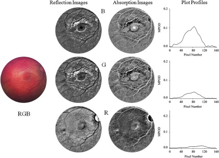
Fig. 3.

Fundus imaging of 4-month-old infant with white-light excitation. Left: raw image obtained with combined RGB-chip readouts of CCD detector array. First column: reflection images derived from raw image via selective B, G, and R-chip readouts, respectively. Second column: B, G, and R absorption images derived from reflection images. For better visual appearance, all B, G, and R images were FFT filtered (intensity variations removed in small structures up to three pixels, and in large structures averaged over 40 pixel increments). Right column: MP optical density (MPOD) line profiles obtained from raw image B, G, and R chip intensities, respectively, along meridians running horizontally through the center of the macula.
