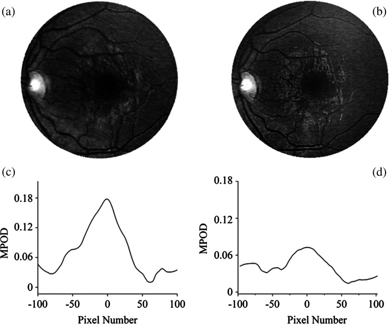
Fig. 5.
Sensitivity of RetCam® imaging to subject’s eye alignment in white-light excitation mode. Examples of gray-scaled B-chip images for slightly different positioning of macula within fundus image (top) and the corresponding line plots along meridians through macula (bottom). Depending on alignment, apparent MPOD results differ by more than a factor or 2 ( versus ).

