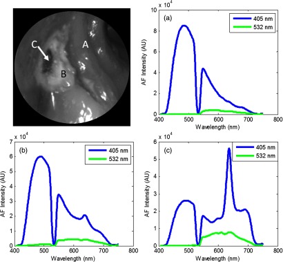
Fig. 12.

Top left: 405-nm reflectance images of an upper-left premolar of a 4-year-old patient with an occlusal lesion. The white spot lesion (B) is evident by the increased reflectance leading to a brighter appearance compared to the surrounding enamel (A). A more severe portion of the lesion (C) is evidenced by the dark spot near the center of the lesion. Top right: AF spectra from a sound region on the tooth (a). Bottom left: AF spectra from the white spot lesion (b). An RPC value of 41% is observed between sound (a) and white spot (b) enamel. Bottom right: AF spectra from the brown spot (c). An RPC value of 70% is observed between sound (a) and lesion (c). Presence of bacterial red fluorescence as evidenced by the spectral feature near 635 nm in both bottom spectra.
