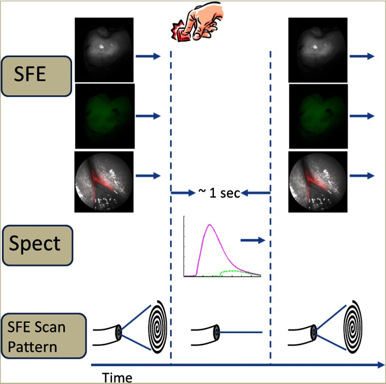
Fig. 3.
The operational flow of the device. The system begins in imaging mode, which captures reflectance, AF, and bacterial fluorescence images. When the clinicians notice a suspicious area, they will center the image over the area and push a software icon button to temporarily disable the imaging mode and begin collecting spectroscopic data. Approximately 1 s later, numerical spectroscopic results are displayed and the system resumes imaging.

