Fig. 4.
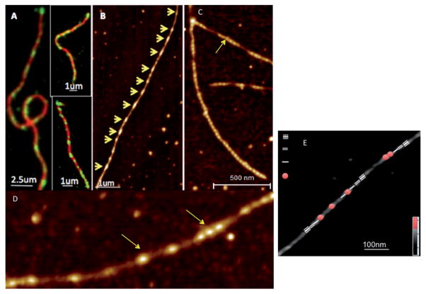
(A) TIRF imaging revealed a cluster-like attachment of drebrin (green) to actin filaments (red), providing the first indications of cooperative interaction between drebrin1–300 and F-actin. (B) AFM images of F-actin decorated with drebrinA1–300 at sub-saturating ratios reveal their cooperative binding (binding clusters are marked by yellow arrows along individual filaments). (C) and (D) show drebrin clusters along the actin filament at higher resolution. (E) Identification of the segments of drebrin free F-actin; 1st, 2nd and 3rd neighbor relative to the drebrin-bound helix in the AFM image of partially decorated Drb1–300–F-actin. Adapted from Sharma S. et al.84 with permission from Cell Press.

