
Fig. 7.
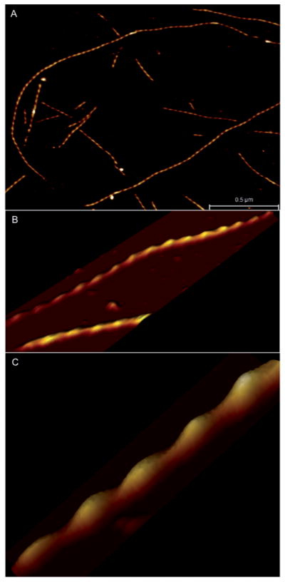
AFM imaging of F-actin under physiological conditions. (A) Image of several actin filaments shows stable attachment to a silane modified mica surface. (B) Higher resolution image of F-actin and (C) 3D view of actin filament section showing well resolved helical periodicity.