Figure 3.
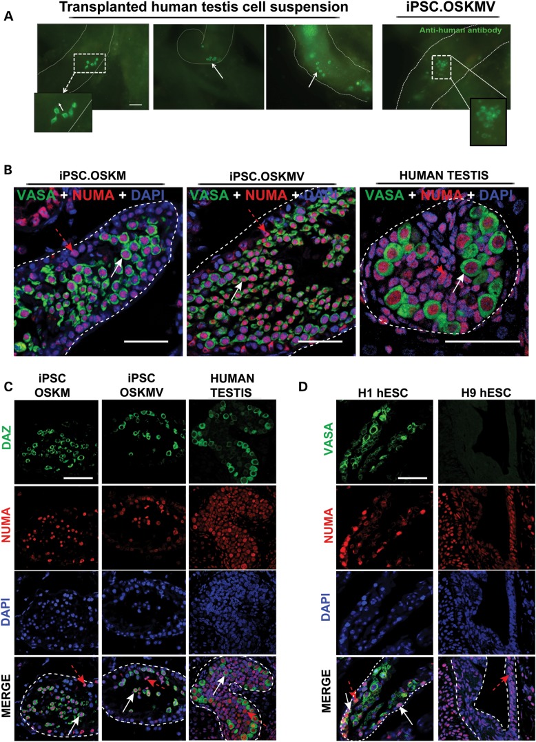
Transplantation of OSKM and OSKMV cells into busulfan-treated mouse testes. (A) Whole-mount analysis on transplanted human fetal testis cells and OSKMV cells into mouse testes. Chain (white dashed rectangle) and cluster formation (white arrows) visible in human fetal testis control cells. Transplanted OSKMV cells gave rise to cluster formation indicated by white dashed rectangle. Scale bar, 50 µm. (B) Histology cross sections of tubules inside mouse testes stained with co-localizing human-specific NUMA (red) and VASA (green). White arrows indicate transplanted cells positive for VASA germ cell marker; red dashed arrows indicate VASA-negative transplanted donor cells. Scale bar, 50 µm. (C) Histology cross sections of tubules of mouse testes stained with additional germ cell-specific marker DAZ demonstrating NUMA co-localization. Scale bar, 50 µm. (D) H1 and H9 hESC controls. H1 cells are positive for VASA/NUMA co-staining; H9 cells are negative for germ cell marker selection. White arrows indicate transplanted cells positive for VASA germ cell marker; red dashed arrows indicate VASA-negative donor cells. Scale bar, 50 µm.

