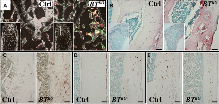
Figure 6.
Dynamic histomorphometric differences and cellular abnormalities in BTKO long bones. (A) In vivo double fluorochrome labeling with calcein (green) and alizarin red shows greatly increased labeled bone surfaces in the trabecular portion of BTKO femur, compared with a control. (B) TRAP staining for osteoclasts (red) is sharply increased in BTKO bone, compared with control, as shown in trabecular portions of the femur. Immunohistochemical staining (brown) performed on cortical femur showed higher levels of MEPE (C) and E-11 (D), and lower levels of sclerostin (E) for BTKO bone than those for controls. Scale bars: 50 µm.

