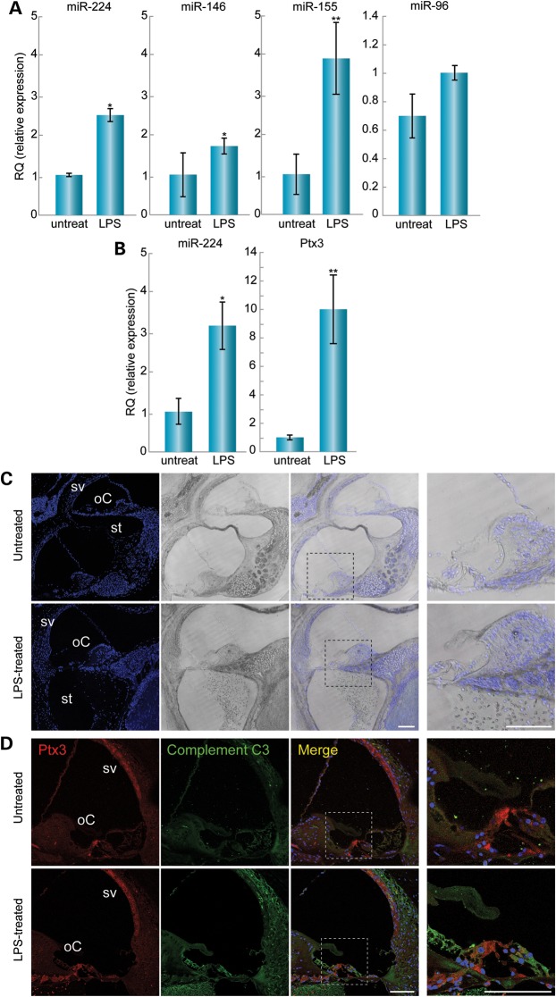
Figure 4.
LPS-induced inflammation in the inner ear leads to changes in RNA expression and activation of the immune system. (A) At 24 h after LPS injection, miR-224 expression was increased by 2.5-fold in the LPS-treated (n = 4) when compared with non-treated (n = 4) ears. *P < 0.05. miR-146 expression was increased by 1.7-fold in the LPS-treated (n = 6) when compared with non-treated (n = 6) ears. *P < 0.05. miR-155 expression was increased by four-fold in the LPS treated (n = 6) when compared with non-treated (n = 6) ears. (**) P < 0.005. There was no significant change in miR-96 expression between the LPS treated (n = 3) and non-treated (n = 3) ears. (B) At 72 h after LPS injection, miR-224 expression was increased by three-fold in the LPS-treated (n = 5) when compared with non-treated (n = 5) ears. (*) P < 0.05. Ptx3 expression was increased by 10-fold in the LPS treated (n = 3) when compared with non-treated (n = 3) ears. (**) P < 0.005. (C) Draq5-stained nuclei in the LPS-treated and non-treated inner ear sections. No morphological changes were observed. Cells were present in the scala tympani in the LPS-treated ears, indicating immune cell infiltration. The inset represents the organ of Corti enlarged for optimal visualization (far right images). Scale bar: 100 µm. (D) At 24 h after LPS injection, Ptx3 staining was observed in the sensory epithelia, mostly in pillar cells in the organ of Corti and in the stria vascularis. Stronger staining was seen in the LPS treated when compared with the non-treated inner ears. Staining with a complement C3 antibody was stronger in the LPS treated when compared with the non-treated inner ears, indicating activation of the immune system. The inset represents the organ of Corti enlarged for optimal visualization (far right images). oC, organ of Corti; sv, stria vascularis. Scale bar: 7.5 µm.

